PHYLUM PORIFERA
The sponges are the first metazoans (multicellular
animals) that we will study. The
principal features of phylum Porifera are listed below.
1. While
some sponges are radially symmetrical, the majority of sponges are asymmetrical
in body form. Sponges are considered to
be at a cellular grade of construction; that is, they have cellular
differentiation (tissues) without cellular coordination.
2. The
outermost tissue layer of sponges is composed of cells called pinacocytes. In some sponges this outer tissue layer is syncytial while in others the
pinacocytes are all distinctly separated from one another by cell
membranes. The innermost tissue layer
is composed of cells called choanocytes
or collar cells (see S&S, p.45) which have flagella that beat to produce
water currents through the sponge body.
Between these two tissue layers is a gelatinous layer called the mesoglea (mesohyl). The mesoglea is not considered to be a
tissue since it contains a number of different kinds of independently
functioning cells. Each cell type in
the mesoglea has a specific name, but the general term for all of these
wandering cells is amoebocyte.
3. Some of
the amoebocytes in the mesoglea are specialized for secreting a skeleton. The sponge skeleton may be composed of
mineral spicules, spongin fibers or a combination of these two, depending on
the kind of sponge. Spicules may be
calcareous (composed of Ca CO3) or siliceous (composed of H2Si2O7). Spongin fibers are composed of a
sulfur-containing schleroprotein.
4. Water
enters the body of a sponge by way of a number of minute incurrent pores or ostia.
Water leaves the body by way of one or more large excurrent pores or oscula. Within the body of the sponge, the water may
pass through a large cavity (the spongocoel)
through a system of canals and chambers, or through a combination of these two.
5. Movement
of water through the sponge body is accomplished by the beating of the
choanocyte flagella. The choanocyte cells
line either a spongocoel or a number of small chambers, depending on the
sponge. A choanocyte cell consists of a
nucleus, one or more vacuoles, a long flagellum and a delicate, collarlike
structure which surrounds the base of the flagellum. Electron microscope studies show the collar
of a choanocyte to be composed of a circular arrangement of microvilli-like
structures extending outward from the cell body. The rotary motion of the flagellum forces
solid food particles in the incoming water to adhere to the outside surface of
the collar. The streaming protoplasm of
the collar transfers the food to the collar base where ingestion can occur.
6. Sponges
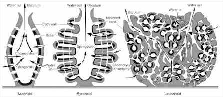
may be divided into three basic grades or types based upon the arrangement of
their water canal systems. Note that
these grades or types are not taxonomic groupings. The three types of sponges are described
below and are shown diagrammatically.
·
Asconoid Type - Water entering the sponge
passes through ostia which are actually openings within doughnut-shaped cells
called porocytes, which are found
only in asconoid sponges. The water
enters the large central cavity called the spongocoel,
which is lined with choanocytes. Water
exits from the spongocoel through a single large osculum.
·
Syconoid Type - Water enters the sponge
through ostia which are openings
between cells, rather than within cells as in asconoid sponges. Water then passes into radially arranged
incurrent canals which lead to flagellated chambers lined with choanocytes. Water leaves the flagellated chambers by way of excurrent canals that lead to the
spongocoel, which is lined a simple flat epithelium. Water exits from the spongocoel by way of a
single large osculum. Note that the
body wall of syconoid sponges is thicker than that of asconoid sponges and that
the syconoid spongocoel is not lined by choanocytes as is the asconoid
spongocoel.
·
Leuconoid Type - The ostia of a leuconoid
sponge are like those of a syconoid sponge.
These ostia lead into a complex system of canals and flagellated
chamgers that penetrate the very thick, dense mesoglea. There is no spongocoel in a leuconoid
sponge. Rather, water reaches the
oscula by way of large excurrent canals.
The complex canal system of leuconoid allows for greater surface area
over which water may pass and consequently creates an increased area for food
and oxygen uptake and for waste removal.
It is not surprising, therefore, that leuconoid sponges are the largest
in size of all the types and that the vast majority of sponges are leuconoid.
7. Sponge
taxonomy is based on skeletal composition.
The four classes in phylun Porifera are listed below along with
distinguishing characteristics for each class.
The grades of sponges found in each class are given in parenthesis,
although this is not distinguishing since there is overlap between the classes.
a) Class Calcarea - contains sponges
having calcareous spicules with 1 to 4 rays.
(asconoid, syconoid, leuconoid)
b) Class Hexactinellida - contains sponges
having siliceous spicules with 6 rays.
These spicules are often fused to form a beautiful lattice-like
cylinder, as in the so-called Venus' flower basket. (syconoid)
c) Class Demospongiae - contains sponges
having siliceous spicules (not 6-rayed) and/or spongin fibers. (leuconoid)
d) Class Sclerospongiae - contains sponges
having an internal skeleton of siliceous spicules and spongin fibers plus an
outer encasement of calcium carbonate.
Only six species from the Caribbean have been described to date. (leuconoid)
8. Sponges
are capable of both sexual and asexual reproduction and they also have great
powers of tissue regeneration and reassociation. Sexual reproduction is
accomplished by production of eggs and sperm which unite to form a zygote. The zygote divides repeatedly to produce a
free-swimming larval form. Depending on
the sponge, this larva may be either a uniformly ciliated parenchymula larva or an amphiblastula
larva, which has flagella only at one pole (Fig. 2.7, p.55 of S&S). The larvae eventually settle and
metamorphose into the adult form.
Sponges may reproduce asexually by budding. In addition, all freshwater sponges and some
marine forms produce resistant overwintering bodies called gemmules. These gemmules
consist of aggregations of food laden amoebocytes surrounded by a resistant
covering. They are produced during
periods of cold or drought and can survive to produce a new sponge body when
conditions improve (Fig. 2.8, p.55 of S&S).
In
today's laboratory you will examine the structure of the sponge species
available and then perform experiments on the reassociation of porifera cells.
ASCONOID SPONGES
Asconoid
sponges are the simplest and most primitive sponge architectural type and are
all relatively small due to their inefficient filtering system. Asconoid structure is demonstrated in Leucosolenia. Obtain a small colony of Leucosolenia sponges and place it in a dish filled with
seawater. Examine it under a dissecting
microscope. Does it respond to a
stimulus? Do you detect movement? To observe the filtering mechanism of Leucosolenia, prepare a dilute
suspension of carmine powder and seawater and then gently place a drop of the
suspension near the colony. Describe
the water movement through Leucosolenia. Where do the carmine particles enter? Where do they exit? Do particles enter the sponge at the same
velocity that they exit? Explain. Do you see budding on your Leucosolenia colony? If so, where are the buds positioned? Examine the colonial structure of Leucosolenia. Describe how you think the ultimate colonial
form develops from a single sponge tube.
From your Leucosolenia colony remove a single
sponge tube for closer examination and then rinse the colony in fresh seawater
and return it to the holding tank. Cut
the sponge tube longitudinally into two halves (from osculum to base) and place
the two halves on a slide so that half the tube shows the inner surface and the
other half shows the outer surface. Add
a drop of saltwater and cover with a coverslip. Examine both surfaces under the compound
microscope. Try to identify porocytes,
pinacocytes, and choanocytes, or evidence of their presence (refer to S&S,
pp. 45‑47 for diagrams). Next, tease apart the sections of sponge
with a dissecting needle and examine for spicules and amoebocytes. Describe the shape and arrangement of the
spicules. How are spicules formed? Do you see any evidence of this in your
preparation?
Examine the prepared slides of asconoid sponges. The staining of these slides will make the
cellular structures easier to identify.


.jpg)


No comments